Throughout the semester I was able to learn about major categories of pathogens: fungi, nematodes, bacteria, and viruses. We focused on the visualization of structures specific to each type of pathogen, the identification process, and management strategies. I was able to learn about common pathogens and their disease triangles. This is a diagram that connects the pathogen to the environment and management practices, allowing for better control of the pathogen in agricultural settings.
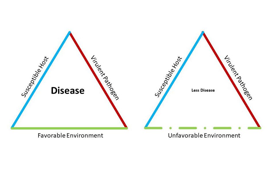
image source: https://www.popularmechanics.com/science/a32238447/disease-triangle-covid-19-coronavirus-flatten-curve/
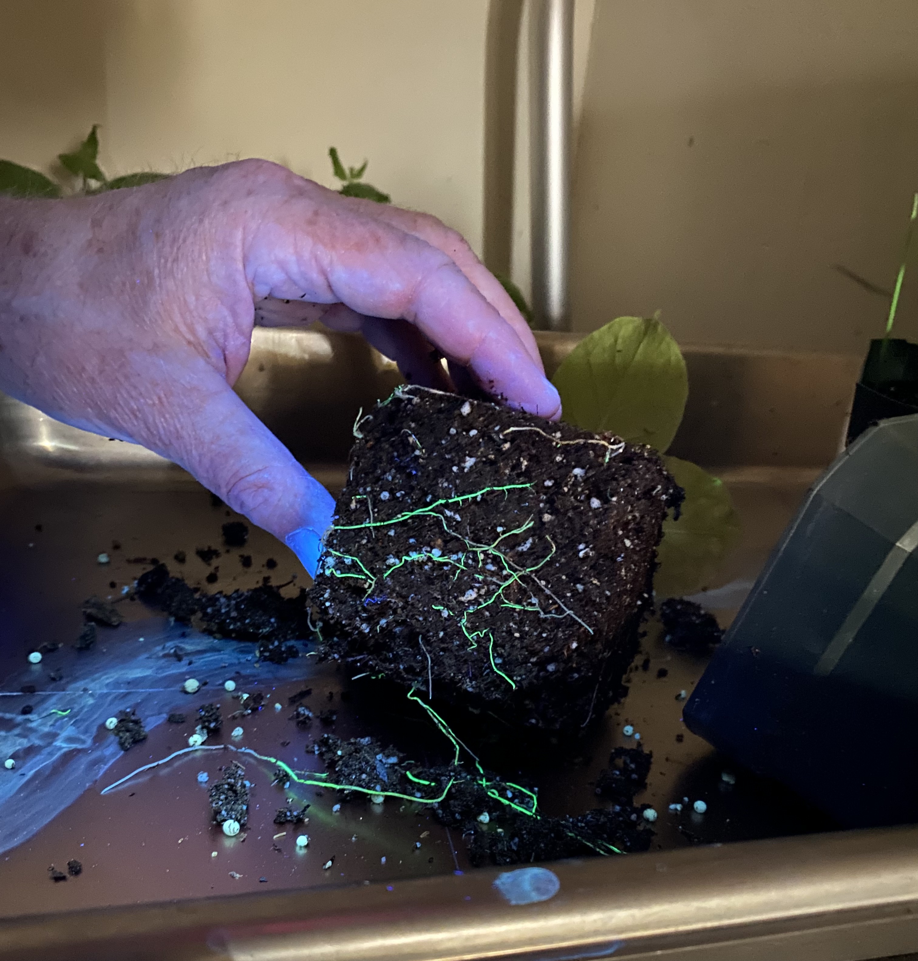
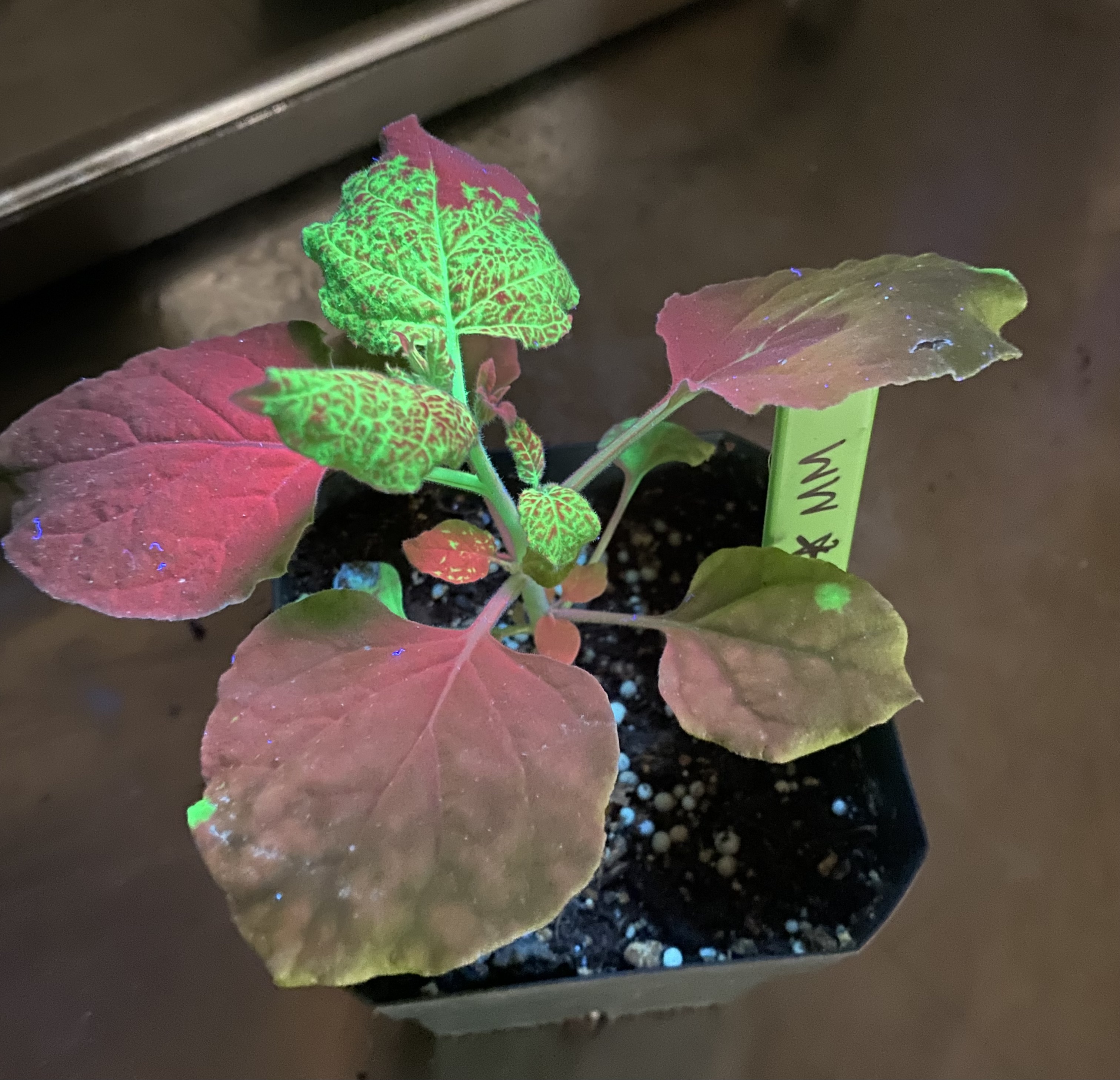
In the lab section of the course, I had the opportunity to run an ELISA test, PCR, try innoculations, and get some more microscopy experience. In the image below, a tobacco plant inoculated with Tobacco Mosaic Virus tagged with a green fluorescent protein (TMV-GFP) can be seen. We were able to visualize the virus’s pattern of movement over the span of a week.
When viruses first invade a plant they move towards the roots and other vascular structures. Then they make the ascent to the top of the plant to colonize young foliar tissues. This is why we can see the TMV-GFP fluorescing under UV light in the roots primarily and partially in the upper shoot of the plant.
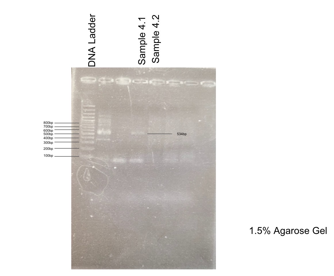
In a separate lab, I was also able to run through the process of PCR. We used this test to determine whether or not our samples contained the Cauliflower Mosiac Virus. Then we were able to run gel electrophoresis to visualize the size of the bands present and make a determination on whether our samples were positive or negative for the virus. We can see in the image below that sample 4.1 was positive and 4.2 was negative.
Overall, I enjoyed every moment in my Plant Pathology class and walked away with new skills and an awareness of how to prevent and manage plant pathogens.